
AI In Microscopy Market Size and Share
Market Overview
| Study Period | 2020 - 2031 |
|---|---|
| Market Size (2026) | USD 1.51 Billion |
| Market Size (2031) | USD 2.98 Billion |
| Growth Rate (2026 - 2031) | 14.53 % |
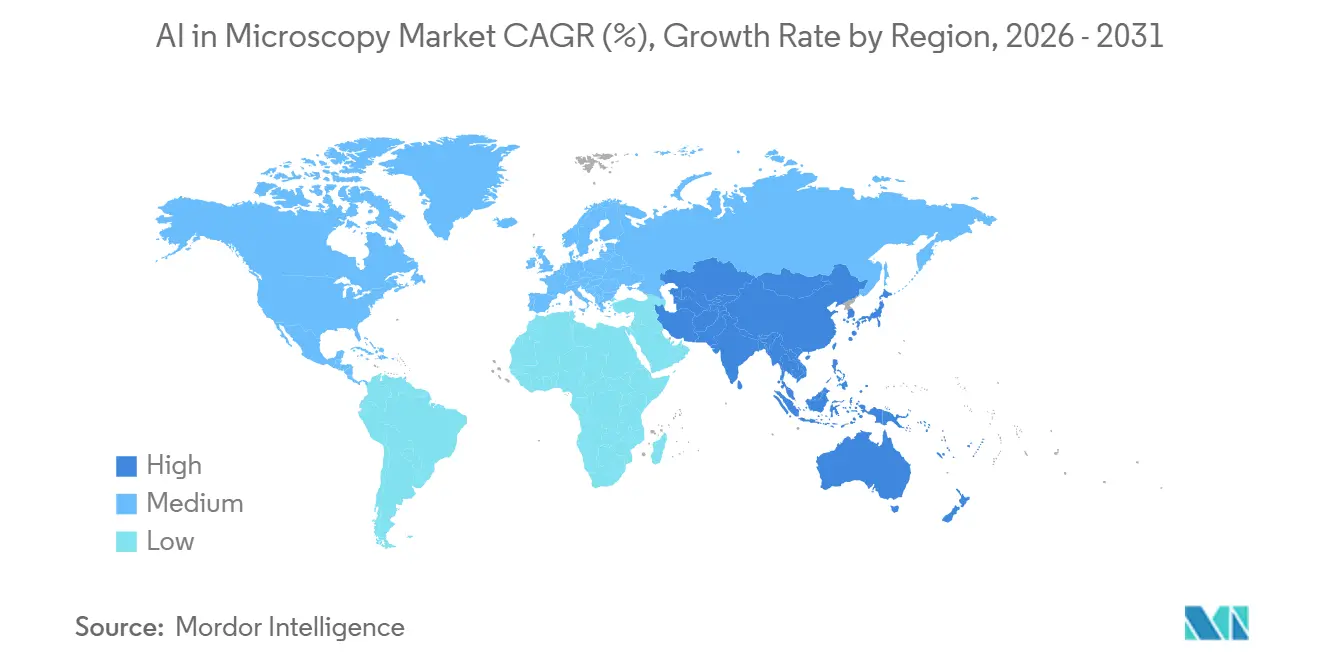
| Fastest Growing Market | Asia-Pacific |
| Largest Market | North America |
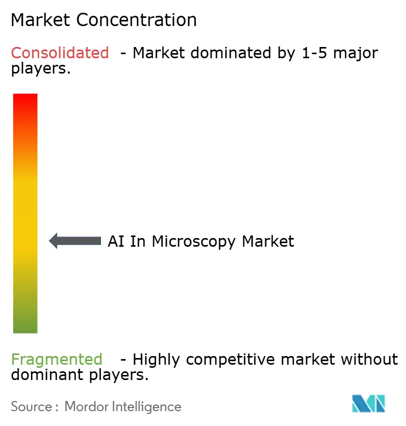
| Market Concentration | Medium |
Major Players *Disclaimer: Major Players sorted in no particular order Image © Mordor Intelligence. Reuse requires attribution under CC BY 4.0. | |
AI In Microscopy Market Analysis by Mordor Intelligence
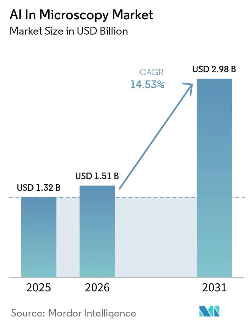
The AI In Microscopy Market size is expected to grow from USD 1.32 billion in 2025 to USD 1.51 billion in 2026 and is forecast to reach USD 2.98 billion by 2031 at 14.53% CAGR over 2026-2031.
The AI microscopy market is moving forward as laboratories shift from manual image review toward digital, software-led workflows that shorten review time and support more consistent interpretation across routine pathology, translational research, and industrial inspection settings. This transition is gaining support from better whole-slide imaging workflows, stronger model training depth, and wider use of AI inside instrument software rather than as a separate add-on. Competitive activity in the AI microscopy market is also rising as large diagnostics and life science companies treat AI pathology and quantitative imaging as core platform capabilities, not optional software features. The strongest opportunities are building around clinical deployment in pathology, high-content imaging in drug development, and instrument-level AI that reduces dependence on external computing and speeds analysis at the point of image capture. The main drag on adoption remains the work needed to validate models across different stains, tissues, scanners, and site-specific workflows, which slows wider procurement even when technical performance is strong.
Key Report Takeaways
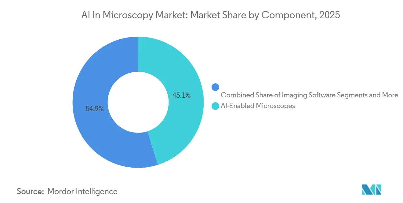
- By component, AI-enabled microscopes held the largest share at 45.1% in 2025, while imaging software is projected to grow fastest at 18.3% CAGR through 2031.
- By technology, deep learning image analysis led with 48.2% of revenue in 2025, while predictive analytics is forecast to expand fastest at 19.2% CAGR through 2031.
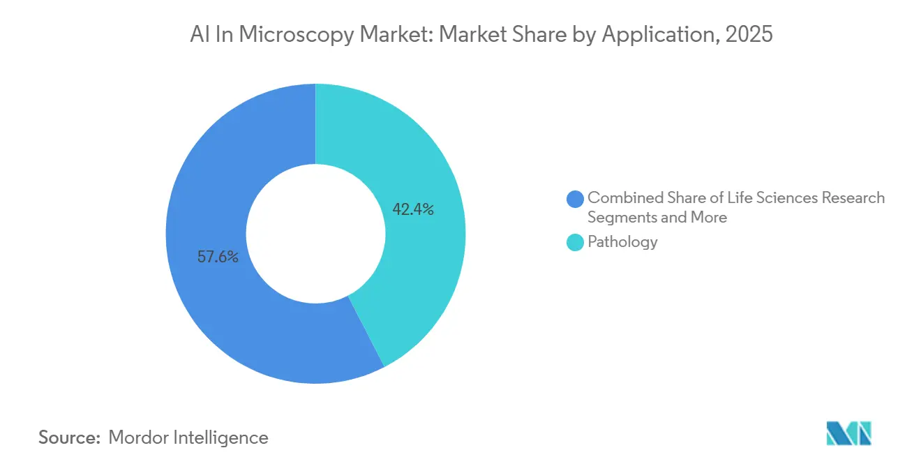
- By application, pathology accounted for 42.4% of revenue in 2025, while drug discovery is expected to record the highest growth at 16.9% CAGR through 2031.
- By end user, hospitals and clinics held the largest share at 44.1% in 2025, while pharmaceutical and biotechnology companies are projected to grow fastest at 18.9% CAGR through 2031.
- By geography, North America led with 40.5% of revenue in 2025, while Asia-Pacific is expected to advance at the fastest pace with a 13.2% CAGR through 2031.
Note: Market size and forecast figures in this report are generated using Mordor Intelligence’s proprietary estimation framework, updated with the latest available data and insights as of January 2026.
Market Trends and Insights
Rising Adoption of AI-Enabled Whole-Slide Imaging in Routine Pathology
Whole-slide imaging is moving from limited pilots into daily use, but the pace still differs by institution and by the maturity of the workflow. A 2026 structured landscape analysis in Virchows Archiv[1] showed that successful clinical deployment depends heavily on fitting AI into existing laboratory information system workflows instead of running it as a separate tool. That finding matters because labs usually buy for workflow fit first, then expand algorithm use after the imaging and case management stack is stable. PathAI’s 2026 deployment agreement with MedStar Health also points to this shift from single-site testing toward broader health system rollouts for digital pathology platforms. Standardized image exchange and tighter links with pathology information systems are making early adoption less disruptive for first-time buyers. The result is that the AI microscopy market is being shaped as much by integration readiness as by raw model accuracy.
Growth of Foundation Models for Cell, Tissue, and Subcellular Image Interpretation
Foundation models are changing how the AI microscopy market builds and scales new applications. Paige’s PRISM2 release showed how a whole-slide model can support multimodal pathology use cases and natural language interaction, which broadens the role of AI beyond narrow single-task output. A University of Cologne team reported in Nature Medicine that the SPARK framework could uncover hidden biological information in routine histology, which pushed AI closer to autonomous discovery in cancer pathology. In 2025, npj Digital Medicine[2] also published PathOrchestra, which was trained on 287,424 whole-slide images across 21 tissue types and delivered strong performance across 47 tasks, including pan-cancer classification and lymphoma subtyping. This kind of model depth reduces the cost of launching new use cases on top of existing image archives and scanner fleets. It also makes proprietary clinical data, validation systems, and regulatory readiness more important than algorithm novelty alone.
Expanding High-Content Screening Workflows in Drug Discovery and Translational Research
High-content imaging is becoming a larger growth engine for the AI microscopy market as drug developers need faster and more quantitative screening systems. In 2025, Nature Communications[3] published the HCS-3DX system, which enabled automated single-cell analysis of 3D organoid cultures in high-throughput screening and improved the relevance of preclinical imaging workflows. In 2026, Leica Biosystems expanded work with AstraZeneca and Daiichi Sankyo to scale computational pathology algorithms through an end-to-end imaging and image management stack, which shows that imaging platforms are now tied more directly to precision oncology programs. PathAI’s AIM-MASH AI Assist also became the first AI-powered pathology Drug Development Tool qualified by both EMA and FDA, which raised the standing of AI-generated pathology endpoints in regulated drug programs. This makes AI imaging spend easier to justify inside pharmaceutical development budgets because it supports both research throughput and submission strategy. It also creates a wider indirect demand channel as contract research organizations standardize on AI-capable imaging systems for multiple sponsors.
Increased Use of Edge AI for Real-Time Microscopy at the Instrument Level
Edge processing is opening another layer of opportunity for the AI microscopy market by moving inference closer to the microscope itself. In 2025, research published in Electronics[4] showed that the HAILO-8L accelerator could outperform cloud-linked GPU pipelines in real-time microscopy classification on embedded hardware at more than 60 frames per second. Nikon’s 2026 launch of the ECLIPSE LV100AMS brought motorized control, automated imaging, and AI-powered defect analysis into one industrial microscope, which reduced the need for external computing in manufacturing workflows. Thermo Fisher Scientific’s collaboration with NVIDIA adds to the same direction by connecting AI models, agents, and scientific instruments more tightly in laboratory operations at Thermo Fisher Scientific. As this model spreads, recurring value shifts toward embedded software, faster decision loops, and more selective use of centralized storage.
Restraints Impact Analysis of AI In Microscopy Market*
| Restraint | (~) % Impact on CAGR Forecast | Geographic Relevance | Impact Timeline |
|---|---|---|---|
| Limited Clinical Validation Across Diverse Stains, Tissues, And Imaging Protocols | -1.8% | Global, strongest in North America and EU regulatory settings | Short term (≤ 2 years) |
| High Data Standardization And Annotation Burden For Model Training | -1.4% | Global, with structural data-access barriers in China and India | Medium term (2-4 years) |
| Cybersecurity And Data Residency Concerns For Cloud-Connected Microscopy | -0.9% | North America, the EU, and MEA | Medium term (2-4 years) |
| Budget Pressure From Instrument Refresh Cycles And Fragmented Lab IT Procurement | -1.1% | Global, most pronounced in emerging markets | Long term (≥ 4 years) |
| Source: Mordor Intelligence | |||
Limited Clinical Validation Across Diverse Stains, Tissues, and Imaging Protocols
Validation remains one of the clearest limits on how fast the AI microscopy market can turn interest into deployed revenue. The core issue is that pathology tools must perform across different stain types, scanner models, tissue handling methods, and local laboratory workflows, which makes evidence generation more demanding than in many other imaging settings. A 2025 review in the Journal of Applied Clinical Microscopy noted that models trained on large academic cohorts often lose accuracy in community settings where staining and calibration vary more widely. Procurement committees are therefore placing more weight on multi-site clinical evidence and workflow readiness than on isolated performance claims. This slows adoption for vendors that have strong algorithms but thin validation depth.
High Data Standardization and Annotation Burden for Model Training
The AI microscopy market also faces a heavy data preparation burden that is hard to remove quickly. Training a clinical model for a single indication can require very large sets of expert annotations, and that process still depends on scarce specialist time. A 2026 study in the Journal of Pathology Informatics described an adoption paradox in which the institutions moving fastest on AI also create proprietary training advantages that strengthen incumbent vendors and raise the barrier for later entrants. Work from Shanghai Jiao Tong University highlighted how hospital data silos in China limit the ability to share training material across institutions, even when installed demand is rising. Foundation models ease part of this problem for common image types, but rare diseases, non-H&E stains, and new biomarker combinations still need targeted annotation. Broader laboratory connectivity may help over time, but data quality and labeling depth will remain a major filter on who can scale credible products.
*Our forecasts treat driver/restraint impacts as directional, not additive. The impact forecasts reflect baseline growth, mix effects, and variable interactions.
AI In Microscopy Market Segment Analysis
By Component:
Hardware Anchors Volume; Software Captures Future ValueAI-enabled microscopes held 45.1% of revenue in 2025, which shows how strongly procurement still starts with core equipment rather than with standalone analytics licenses. In many hospitals and laboratories, the hardware purchase fixes the workflow for years because AI capability is bundled into an asset that remains in use through a long refresh cycle. That pattern keeps the installed base important in the AI microscopy market, especially where buyers prefer integrated systems over assembling separate tools from different vendors. Digital slide scanners also remain central because they convert legacy glass workflows into digital image pipelines that can support storage, sharing, and later model deployment. Storage and archiving solutions benefit from rising whole-slide image volumes. Still, their value mix is changing as faster local inference reduces the share of images that always need centralized cloud processing. This means hardware still anchors current revenue, but it does not fully capture where future value is moving.
Imaging software is the fastest-growing component with an 18.3% CAGR through 2031, which reflects how new analytical features can be added to existing scanner fleets without full capital replacement. In this section of the AI microscopy industry, software growth is tied to model updates, workflow integration, and better user access rather than to new physical installation alone. ZEISS has leaned into that shift with its ZEN AI Toolkit and open model approach, which makes the instrument software layer a practical distribution path for third-party or customer-trained models. That approach matters because buyers increasingly want analytical freedom on top of scanners they already own. The AI microscopy market share held by hardware remains large today. Still, software is where margin expansion is easier because value can be added through updates, model deployment, and workflow tools rather than through another full instrument sale. The result is a market in which hardware secures installed presence while software increasingly shapes recurring revenue and customer lock-in.
By Technology:
Deep Learning Dominant; Predictive Analytics Redefines the Value PropositionDeep learning image analysis held 48.2% of technology revenue in 2025, which confirms that convolutional and transformer-based methods remain the main engine behind current deployment. These models are already embedded across digital pathology, high-content imaging, and industrial defect inspection, so they form the base layer of the AI microscopy market. Their current strength comes from image interpretation at scale, where they support detection, classification, and feature extraction in settings that would otherwise depend on manual review. This dominance also reflects the fact that most commercial systems still sell clear, task-based performance to labs that want immediate operational gains. In simple terms, deep learning remains the default technology buyers encounter first.
Predictive analytics is the fastest-growing technology with a 19.2% CAGR through 2031 because customers increasingly want tools that move from identifying what is visible to estimating what is likely to happen next. Pharmaceutical and biotechnology users are especially important here because they need models that help with response prediction, biomarker-linked stratification, and trial design. Bruker’s 2026 addition of 3D AI cell segmentation models to its AtoMx Spatial Informatics Platform also shows how segmentation, classification, and downstream prediction are being connected into one workflow rather than treated as isolated steps. In industrial use, automated detection remains important, as shown by published work around semiconductor image interpretation and defect localization at production speed. The broader change is that the AI microscopy industry is now selling continuous analytical pipelines that detect, segment, classify, and predict in a connected process. That is why premium contracts increasingly go to platforms that combine several model types inside one environment.
By Application:
Pathology Leads Revenue; Drug Discovery Disrupts the Value ChainPathology held 42.4% of application revenue in 2025, giving it the largest position across the AI microscopy market because it sits at the center of whole-slide imaging adoption, laboratory workflow digitization, and clinical deployment. Health systems continue to treat pathology as the first large-scale setting where digital imaging and AI can produce operational value without changing the full care pathway at once. PathAI’s 2026 agreement with MedStar Health reflects that shift from pilot work toward broader network deployment of image management and AI-supported pathology. Pathology also benefits from strong image volume, repetitive review tasks, and clear workflow pressure on specialist staff. For these reasons, the application base of the AI microscopy market still begins with pathology.
Drug discovery is the fastest-growing application with a 16.9% CAGR through 2031 because pharmaceutical companies are using imaging not only for visualization, but also for regulated evidence generation and higher-throughput screening. PathAI’s dual EMA and FDA qualification for AIM-MASH AI Assist gave drug developers a stronger reason to fund AI histopathology tools as part of submission planning rather than only as research support. In parallel, 2025 work on the ViTally Consistent model showed how large microscopy training sets can improve the quantitative use of cell imaging in life sciences research. Material science is also becoming more important as AI-guided image analysis reaches production-level quality control in semiconductor workflows. The AI microscopy market size tied to pathology is still larger today. Still, the fastest change in spending is happening where imaging links directly to drug screening, trial design, and translational workflows. That changes how vendors package value because pharmaceutical buyers are more willing to pay for throughput, reproducibility, and evidence support than for image capture alone.
By End User:
Hospitals Drive Volume; Pharma and Biotech Lead GrowthHospitals and clinics held 44.1% of end-user revenue in 2025, which puts them at the center of current deployment volume in the AI microscopy market. Their lead comes from routine pathology case loads, growing acceptance of digital pathology infrastructure, and the practical need to improve productivity in specialist review workflows. These buyers usually move carefully, but once a system is selected, it can spread across multiple labs and departments over time. The end-user mix, therefore, reflects volume-led adoption in large care settings rather than fast experimentation alone. Hospitals remain the broadest installed base for current revenue generation.
Pharmaceutical and biotechnology companies are the fastest-growing end-user group with an 18.9% CAGR through 2031 because they combine high-content screening, companion diagnostic work, and clinical image analysis under one spending logic. In this part of the AI microscopy industry, buyers often have clearer budget ownership for software and data tools than hospitals do, which lets deployment move faster. Diagnostic laboratories also play a solid role because large batch volumes improve the economics of AI-assisted review and reduce per-slide analysis cost. Research and academic institutes shape product direction by generating validation evidence, testing new modalities, and helping vendors prove broader clinical relevance. Contract research organizations add another important layer because they purchase imaging capacity on behalf of multiple pharmaceutical programs and create a derived demand channel.
Geography Analysis
North America AI in Microscopy Market
North America held 40.5% of revenue in 2025, which gave it the largest regional position in the AI microscopy market. The region benefits from a dense base of academic medical centers, strong pathology software activity, and faster integration of digital imaging into laboratory workflows. Enterprise deployment interest remains centered on pathology platforms that can handle image management, review, and AI use in the same environment.
Europe AI in Microscopy Market
Europe continues to hold a meaningful share of the AI microscopy market because it combines strong optical engineering capability with established translational research infrastructure. The AI microscopy market share in Europe also benefits from buyers who place high value on technical quality, interoperability, and compliance-ready workflows.
APAC AI in Microscopy Market
Asia-Pacific is the fastest-growing region with a 13.2% CAGR through 2031, which makes it the most dynamic geography in the AI microscopy market. Growth is being supported by laboratory modernization, expanding hospital capacity, and faster movement toward digital imaging in pathology and industrial inspection. Japan added to that momentum in 2026 when PHC received Class II medical device approval for the Ephredia E1000 Dx digital pathology system, and Sectra launched Japan’s first digital pathology project at Kameda Medical Center. South Korea also strengthens the regional profile because semiconductor manufacturing supports demand for AI-powered microscopy in production workflows. The AI microscopy market is therefore geographically led by North America today, while Asia-Pacific provides the clearest runway for long-term expansion.
Competitive Landscape
The AI in microscopy market remains moderately fragmented. The AI microscopy market includes global optical and instrument companies, AI-native pathology software firms, and hybrid spatial biology or workflow platforms that sit between those two groups.
Large hardware participants include Carl Zeiss AG, Leica Microsystems GmbH, Nikon, Evident, Thermo Fisher Scientific, and Hamamatsu Photonics. In contrast, software-led specialists include PathAI, Paige, Visiopharm, Indica Labs, and Aiforia Technologies. Hybrid and adjacent platform players such as Bruker, 3DHISTECH, OptraSCAN, and Sectra add another layer of competition by combining imaging, informatics, and analytical workflows. Competition differs by layer. Hardware concentration is moderate because a small group of major instrument suppliers controls a large share of installed systems, while software and services remain far more fragmented. ZEISS, Nikon, and Leica have all pushed toward software-centered platform strategies where the microscope is part of a wider analytical environment rather than the full value proposition. Open model policies matter in this setting because they turn installed instruments into delivery channels for new AI applications. Vendors that can support external models, workflow integration, and easier deployment are positioned more strongly than those that rely only on closed hardware strength.
The competitive outcome is likely to favor companies that combine validation depth, installed workflow presence, and scalable software attachment. Pure algorithm providers can still win in niche areas, but they face more pressure than before unless they control differentiated data or regulatory pathways. The AI microscopy market is therefore best described as moderately concentrated in hardware and fragmented in software, with consolidation rising around clinically relevant platform assets.
AI In Microscopy Industry Leaders
-
Carl Zeiss AG
-
Leica Microsystems GmbH
-
Nikon Corporation
-
Evident Corporation
-
Hamamatsu Photonics K.K.
- *Disclaimer: Major Players sorted in no particular order
AI In Microscopy Market Companies Covered in this Report
- 3DHistech
- Aiforia Technologies Oyj
- Applied Materials Inc.
- Bio-Rad Laboratories
- Bruker
- Carl Zeiss
- Evident Corporation
- Hamamatsu Photonics
- Indica Labs, Inc.
- Danaher
- Nikon
- OptraSCAN, Inc.
- Oxford Instruments
- Paige.AI, Inc.
- PathAI, Inc.
- PerkinElmer
- Roche
- Sectra
- Thermo Fisher Scientific
- Visiopharm
Recent Industry Developments in AI In Microscopy Market
- May 2026: Roche entered a definitive merger agreement to acquire PathAI for USD 750 million upfront, plus up to USD 300 million in milestone payments. The transaction targets PathAI's AISight Image Management System and companion diagnostic algorithm pipeline.
- April 2026: ZEISS Research Microscopy Solutions and EDGE Biotechnologies announced a strategic collaboration to integrate EDGE's AI-accelerated quantitative imaging assays into ZEISS's hardware and analytics ecosystem.
- April 2026: PathAI and MedStar Health announced a multi-year strategic collaboration to deploy the AISight Dx digital pathology platform across MedStar's multi-site health system, including participation in PathAI's Precision Pathology Network for joint research and AI diagnostic co-development.
- March 2026: PathAI received US FDA Breakthrough Device Designation for PathAssist Derm, an AI solution for whole-slide image analysis of skin lesions in dermatopathology, adding to its portfolio of AISight Dx and AIM-MASH AI Assist.
Global AI In Microscopy Market Report Scope
As per the scope of the market, AI in Microscopy refers to the application of artificial intelligence algorithms to analyze, enhance, and interpret microscopic images, enabling automated detection, classification, and quantification of biological or material structures with greater speed, accuracy, and reproducibility.
The AI in Ophthalmology Market Report segments the market by component, including AI-enabled microscopes, imaging software, digital slide scanners, and storage and archiving solutions. It also categorizes the market by technology, covering deep learning image analysis, automated detection, predictive analytics, and image segmentation and classification models. The application segmentation includes pathology, life sciences research, material science, and drug discovery. The end-user segmentation includes hospitals and clinics, diagnostic laboratories, research and academic institutes, pharmaceutical and biotechnology companies, and other end users. Geographically, the market is segmented into North America, Europe, Asia-Pacific, the Middle East & Africa, and South America. The market report also covers the estimated market sizes and trends for 17 countries across major regions globally. For each segment, the market size and forecast are provided in terms of value (USD).
| AI-Enabled Microscopes |
| Imaging Software |
| Digital Slide Scanners |
| Storage and Archiving Solutions |
| Deep Learning Image Analysis |
| Automated Detection |
| Predictive Analytics |
| Image Segmentation and Classification Models |
| Pathology |
| Life Sciences Research |
| Material Science |
| Drug Discovery |
| Hospitals and Clinics |
| Diagnostic Laboratories |
| Research and Academic Institutes |
| Pharmaceutical and Biotechnology Companies |
| Other End User (Contract Research Organization and Government Agencies, among others) |
| North America | United States |
| Canada | |
| Mexico | |
| Europe | Germany |
| United Kingdom | |
| France | |
| Italy | |
| Spain | |
| Rest of Europe | |
| Asia-Pacific | China |
| India | |
| Japan | |
| Australia | |
| South Korea | |
| Rest of Asia-Pacific | |
| Middle East and Africa | GCC |
| South Africa | |
| Rest of Middle East and Africa | |
| South America | Brazil |
| Argentina | |
| Rest of South America |
| By Component | AI-Enabled Microscopes | |
| Imaging Software | ||
| Digital Slide Scanners | ||
| Storage and Archiving Solutions | ||
| By Technology | Deep Learning Image Analysis | |
| Automated Detection | ||
| Predictive Analytics | ||
| Image Segmentation and Classification Models | ||
| By Application | Pathology | |
| Life Sciences Research | ||
| Material Science | ||
| Drug Discovery | ||
| By End User | Hospitals and Clinics | |
| Diagnostic Laboratories | ||
| Research and Academic Institutes | ||
| Pharmaceutical and Biotechnology Companies | ||
| Other End User (Contract Research Organization and Government Agencies, among others) | ||
| By Geography | North America | United States |
| Canada | ||
| Mexico | ||
| Europe | Germany | |
| United Kingdom | ||
| France | ||
| Italy | ||
| Spain | ||
| Rest of Europe | ||
| Asia-Pacific | China | |
| India | ||
| Japan | ||
| Australia | ||
| South Korea | ||
| Rest of Asia-Pacific | ||
| Middle East and Africa | GCC | |
| South Africa | ||
| Rest of Middle East and Africa | ||
| South America | Brazil | |
| Argentina | ||
| Rest of South America | ||
Key Questions Answered in the Report
What is the expected value of AI microscopy by 2031?
The AI microscopy market is forecast to reach USD 2.98 billion by 2031, rising from USD 1.51 billion in 2026 at a 14.5% CAGR over 2026-2031.
Which region leads global revenue today?
North America led with 40.5% of global revenue in 2025, supported by strong pathology infrastructure, academic medical centers, and enterprise deployment activity.
Which region is growing the fastest through 2031?
Asia-Pacific is projected to grow the fastest at a 13.2% CAGR through 2031, helped by laboratory modernization, hospital expansion, and rising digital pathology adoption.
Which component is expanding the fastest?
Imaging software is the fastest-growing component, with an 18.3% CAGR through 2031, because new analytical capability can be added on top of existing scanner infrastructure.
Why is drug discovery becoming more important in this field?
Drug discovery is growing at 16.9% CAGR because AI imaging now supports high-content screening, translational workflows, and even regulated evidence generation in drug programs.
What is the biggest barrier to broader adoption?
The biggest barrier is still validation across different stains, tissues, scanners, and site-specific workflows, which makes scale-up slower even when algorithms perform well.